‘…[P]erhaps I ought to explain,’ added the badger, lowering his papers nervously and looking at Wart over the top of them, ‘that all embryos look very much the same. They are what you are before you are born – and, whether you are going to be a tadpole or a peacock or a cameleopard or a man, when you are an embryo you just look like a peculiarly repulsive and helpless human being. I continue as follows:
The embryos stood in front of God, with their feeble hands clasped politely over their stomachs and their heavy heads hanging down respectfully, and God addressed them.
He said: “Now, you embryos, here you are, all looking exactly the same, and We are going to give you the choice of what you want to be. When you grow up you will get bigger anyway, but We are pleased to grant you another gift as well. You may alter any parts of yourselves into anything which you think will be useful to you in later life.’
- T.H. White, The Once and Future King
I have always been passionate about the small world; cells, microbes and the like. I vaguely remember being introduced to microscopy in school and peering down through the microscope at a world that had hitherto remained invisible to me, but where the fundamental basis of all life lay. Seeing the cells of a simple onion peel was enough to get me excited about microscopy. With time, this excitement and intrigue only grew bigger, and has led me to witness incredible sights through the medium of the microscope.
Have you ever wondered how an organism comes to be, practically out of nothing? I am sure I’m not the only one to be fascinated by the phenomenon of creation. I have always wondered about what happens from the time an egg is fertilised to the point where it turns into a specific organism.
Molluscs have been instrumental in developing our understanding of the very fundamental processes of embryology and developmental biology, and the Lymnaea stagnalis, among other freshwater snails, has contributed immensely.
For my MSc. final project at the University of Nottingham, I had worked on a film and a book depicting the embryonic development of the Lymnaea stagnalis. This project involved extensive research, and over a month spent bending over a microscope to create the necessary images through microscopic imaging.
This story is just a teaser of my larger project. It is the journey of the Lymnaea stagnalis, from embryo to complex juvenile adult.
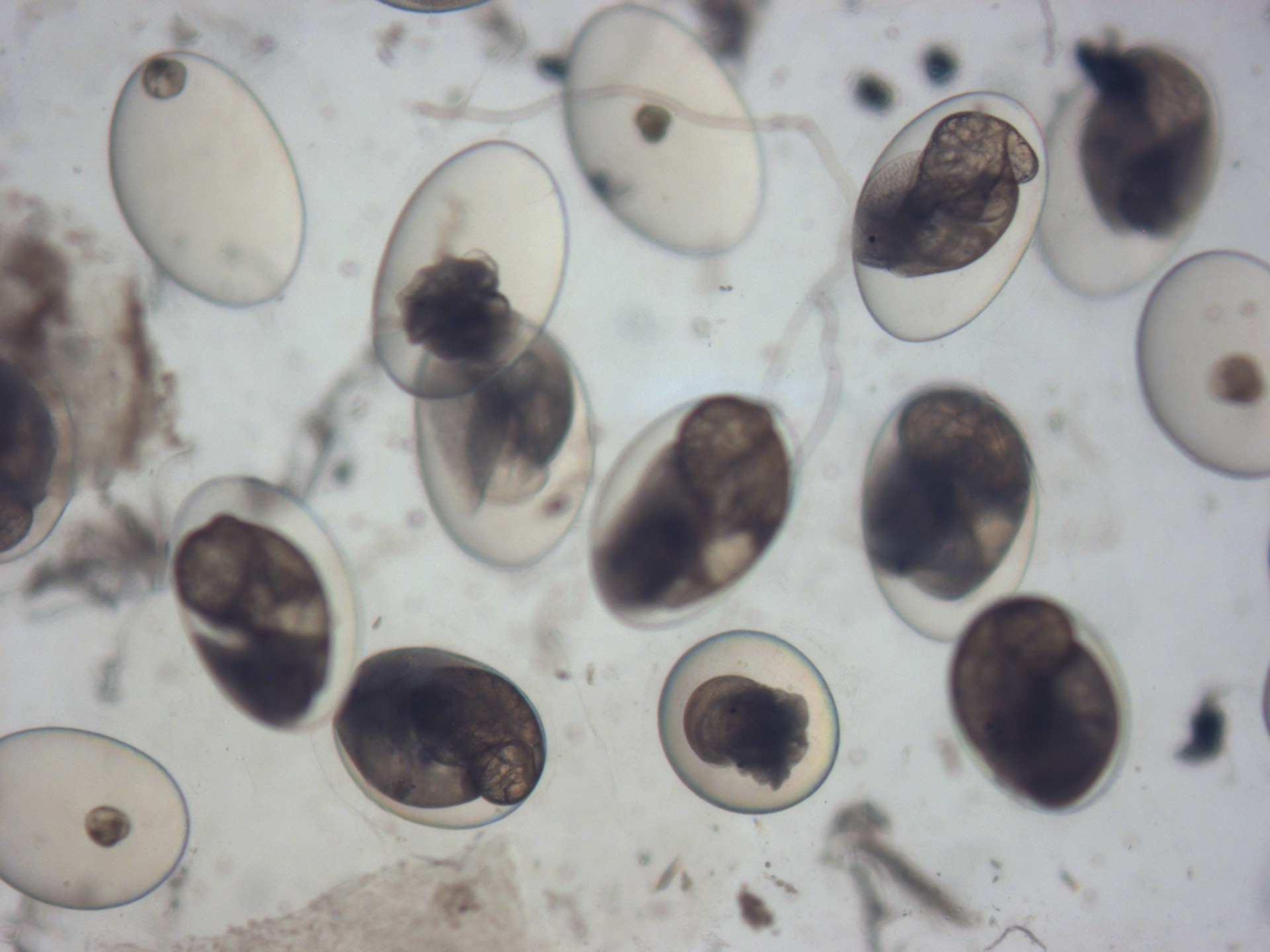
This is a glorious sight of several freshwater pond snails in various stages of embryo development. Every single one of those beautifully transparent egg capsules allow for the observation of growing embryos, while also acting as diffusion barriers that prevent the passage of large molecules. In other words, they do a swell job of keeping the embryos within nourished and safe. (As seen under a stereo microscope.)
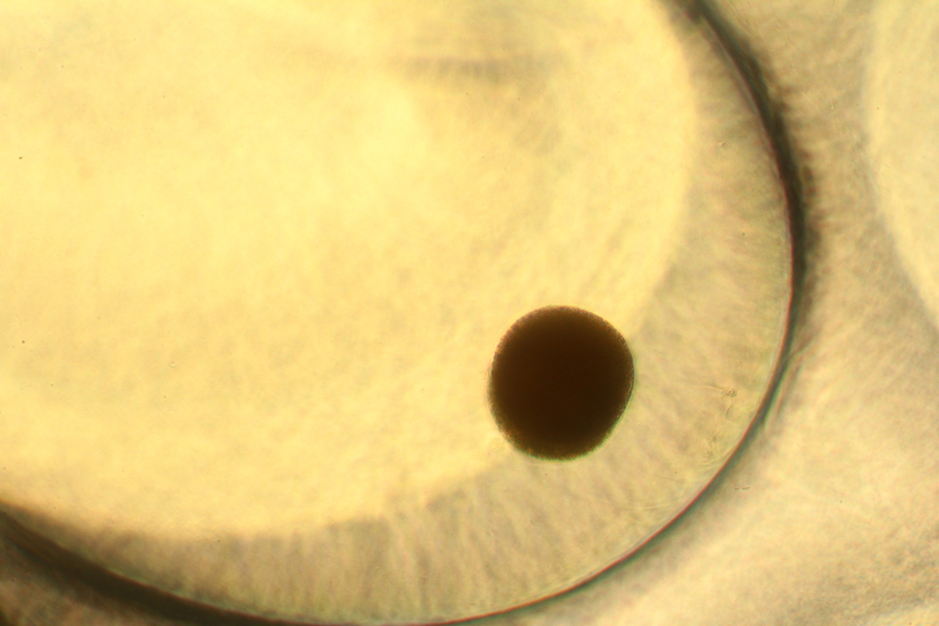
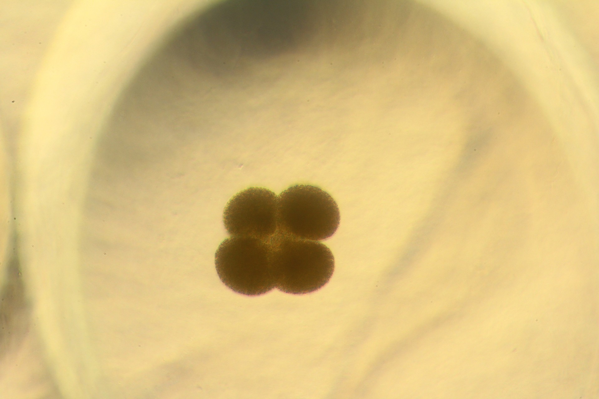
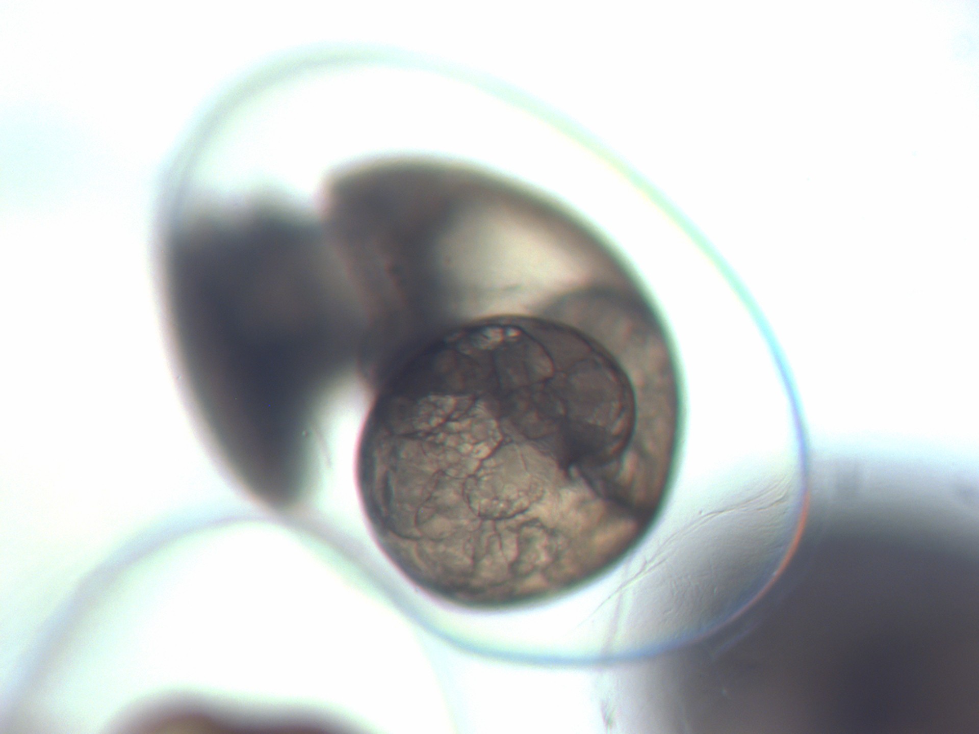
Lymnaea stagnalis lays eggs whose capsules are entirely transparent. This is why it is possible to observe the embryonic development of live specimens under a microscope. The journey starts off with a single-celled fertilised zygote, which almost immediately starts to divide. Several cleavages later, the embryo grows in size and cells in number, till it starts to discernibly acquire a distinct form. (As seen under a compound microscope and magnified 20x.)
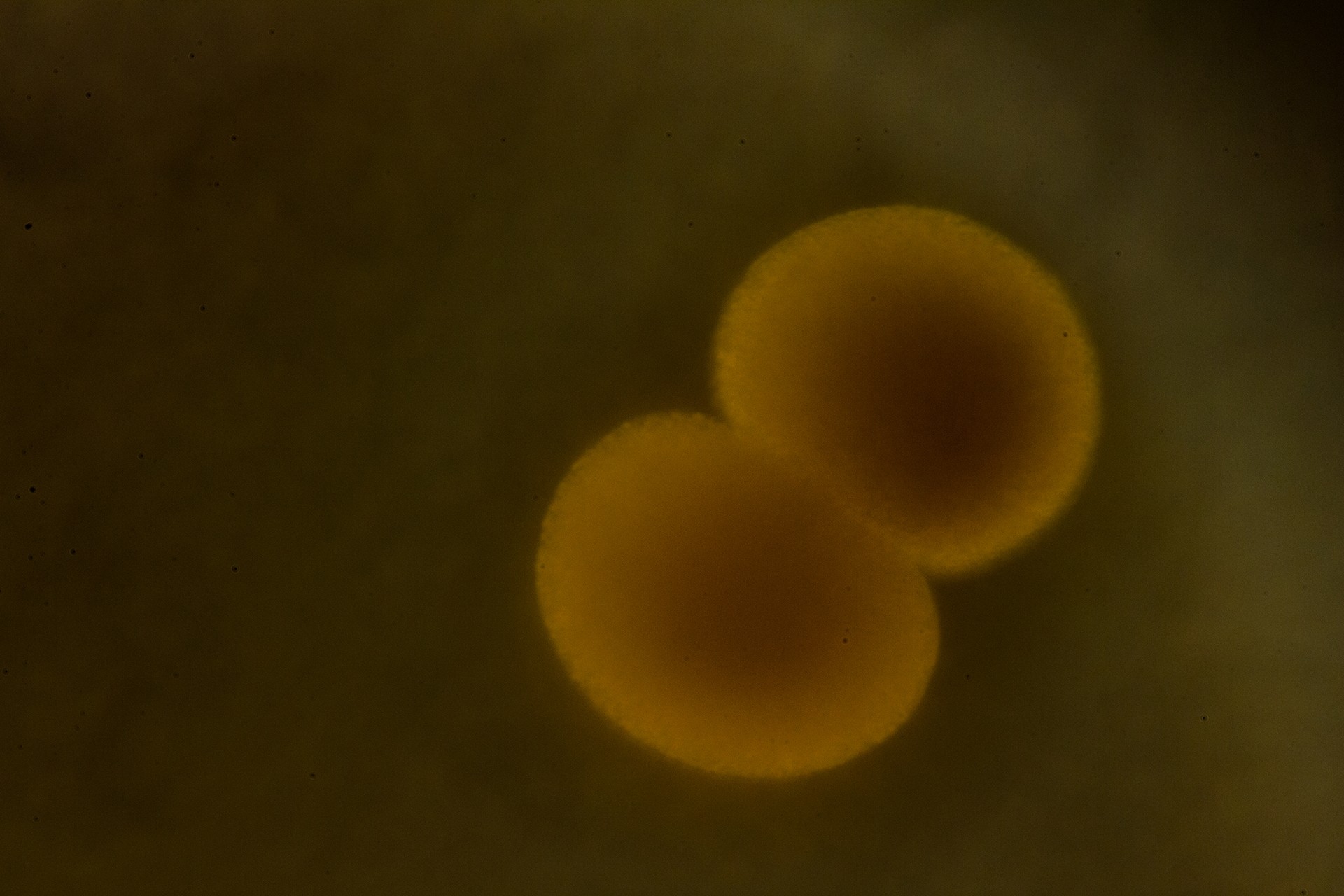
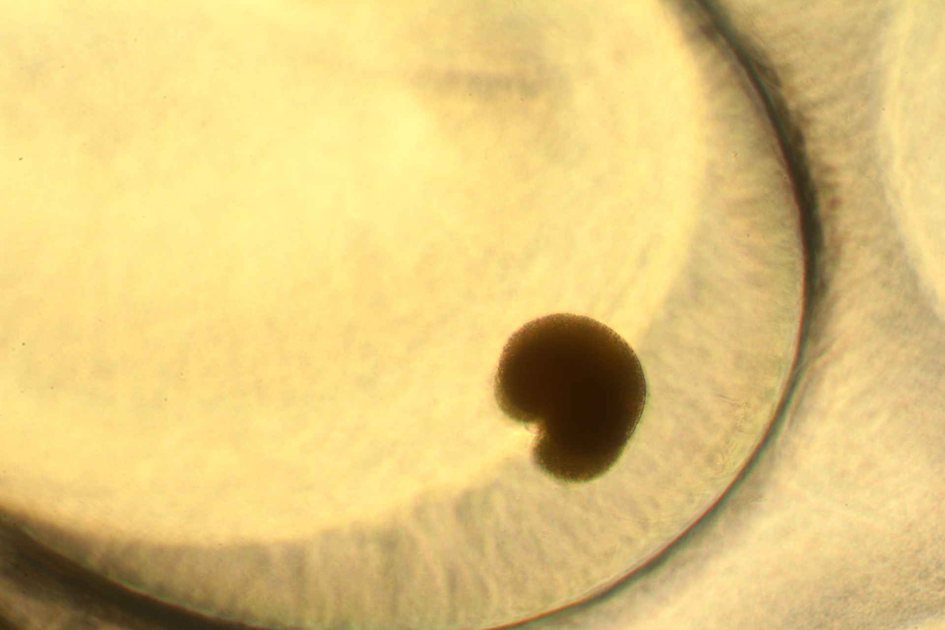
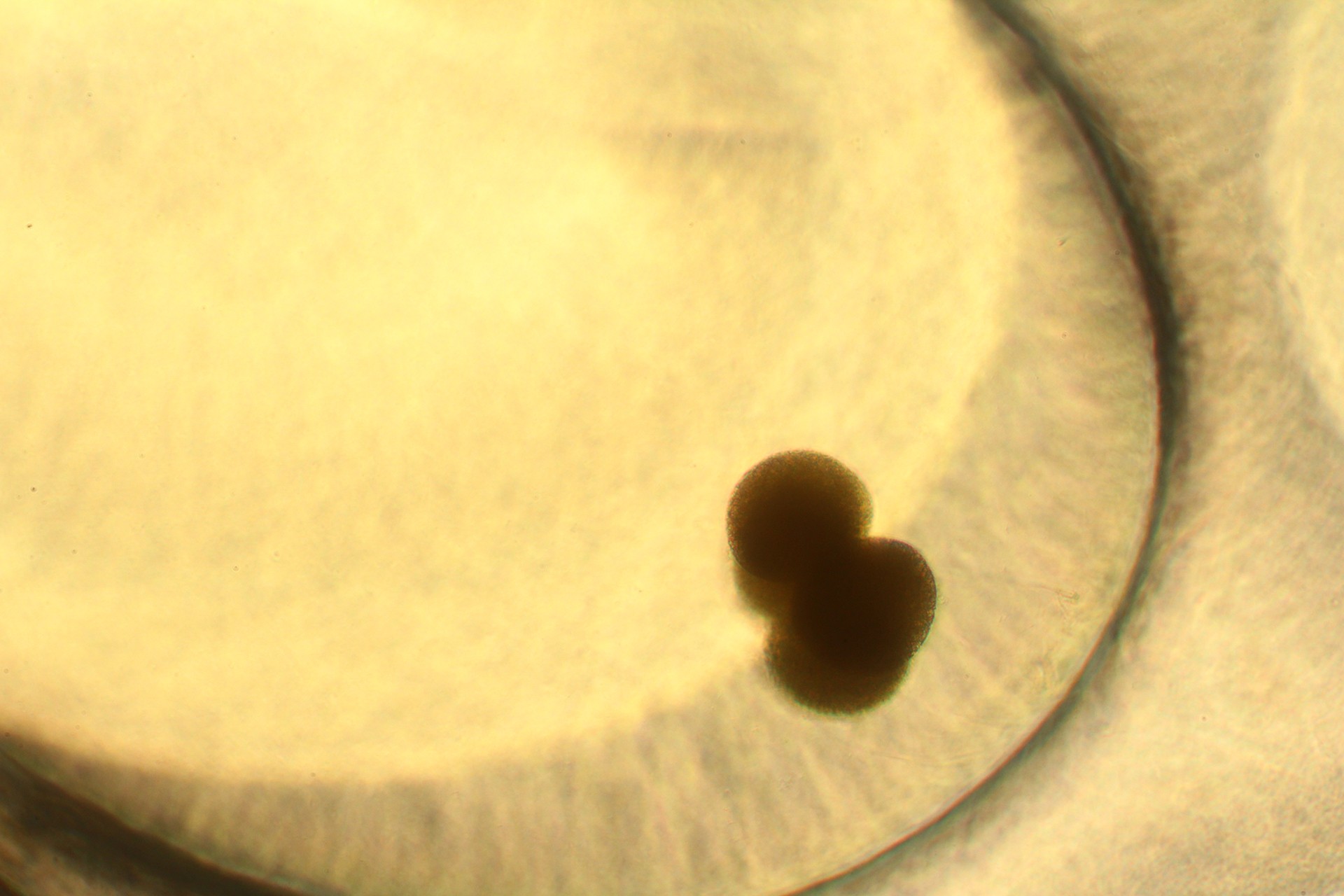
A closer view of a two-celled embryo after the first cleavage. An embryo has so much going on, on the inside and the outside. There are so many factors at play, all coming together for the sole purpose of enabling the embryo to form, and survive. (As seen under a compound microscope and magnified 40x.)
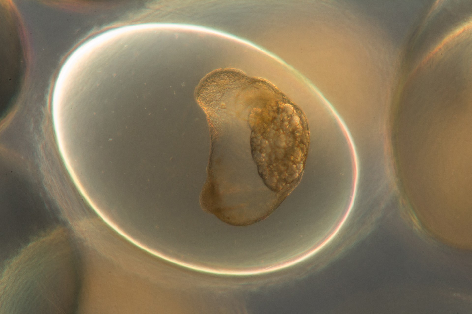
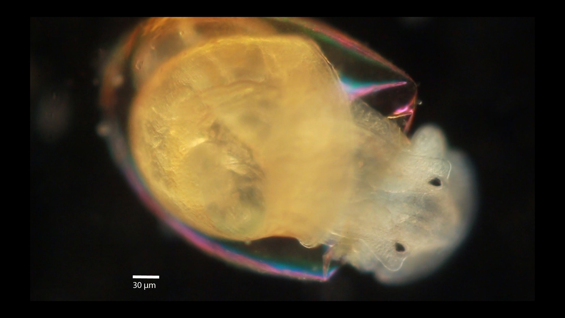
This is the trochophore stage of the snail embryo that develops within two days of the egg being laid. It's when the cells are busy organising and gearing up for more specialised roles; the nutrient-rich fluid within the cell capsule ensuring this growth continues unabated. (As seen under a compound microscope and magnified 10x.)
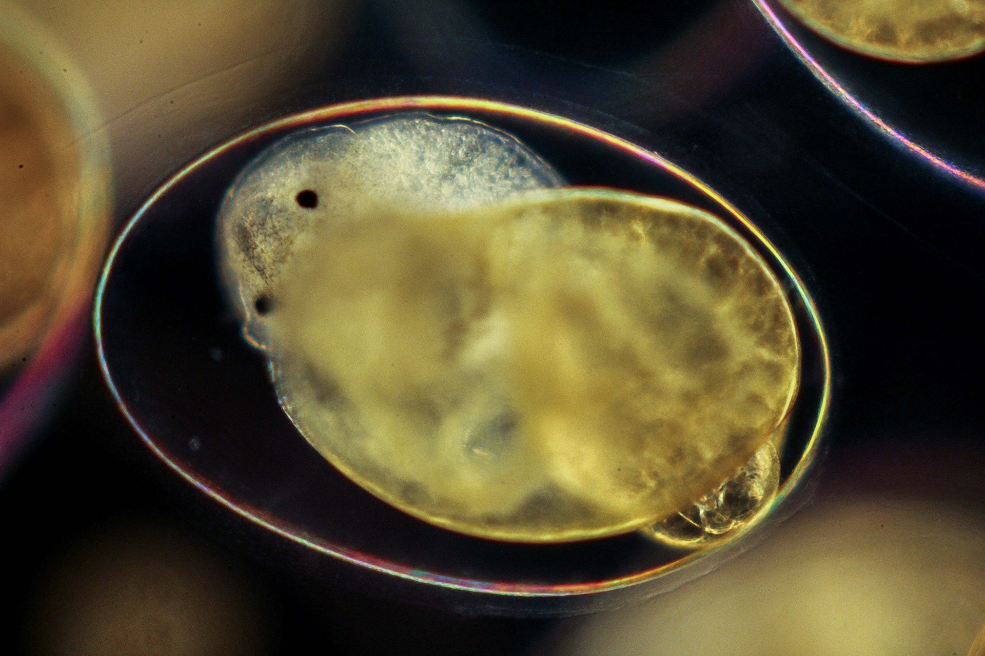
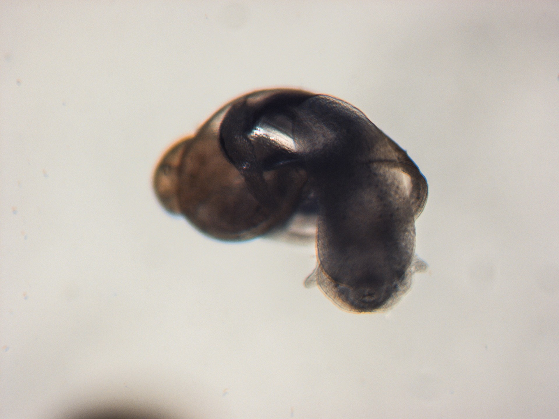
Soon, the embryo develops a bilobed foot, characteristic of a snail. A larval shell is formed and the first hint of asymmetry begins to show in the embryo. After about four days, the embryo enters the metamorphic period and begins to resemble a miniature hippopotamus when seen from the front. That’s why this stage in its larval life is termed the ‘hippo’ stage. (As seen under a compound microscope and magnified 10x.)
Slowly, the embryo metamorphoses into a juvenile adult. The beautiful, distinct snail spiral is now clearly visible. This is marked by the appearance of rudimentary eyes and tentacles. The larval shell covering most of the posterior section and the lung and heart are also slowly developing. (As seen under a stereo microscope.)
Once the primordial organs and tissues become more specialised and differentiate, and all the nutrients within the capsule get exhausted, the juvenile is ready to hatch and make its entry into the world. Here, a juvenile is seen taking its first ‘steps’ into the world. A living being is born! (As seen under a compound microscope and magnified 10x.)
Hardly a millimetre long, the just-emerged juvenile adult Lymnaea stagnalis possesses all the structures of the adult snail. This feat is achieved within a little more than a week’s time since the eggs were first laid. (As seen under a stereo microscope.)
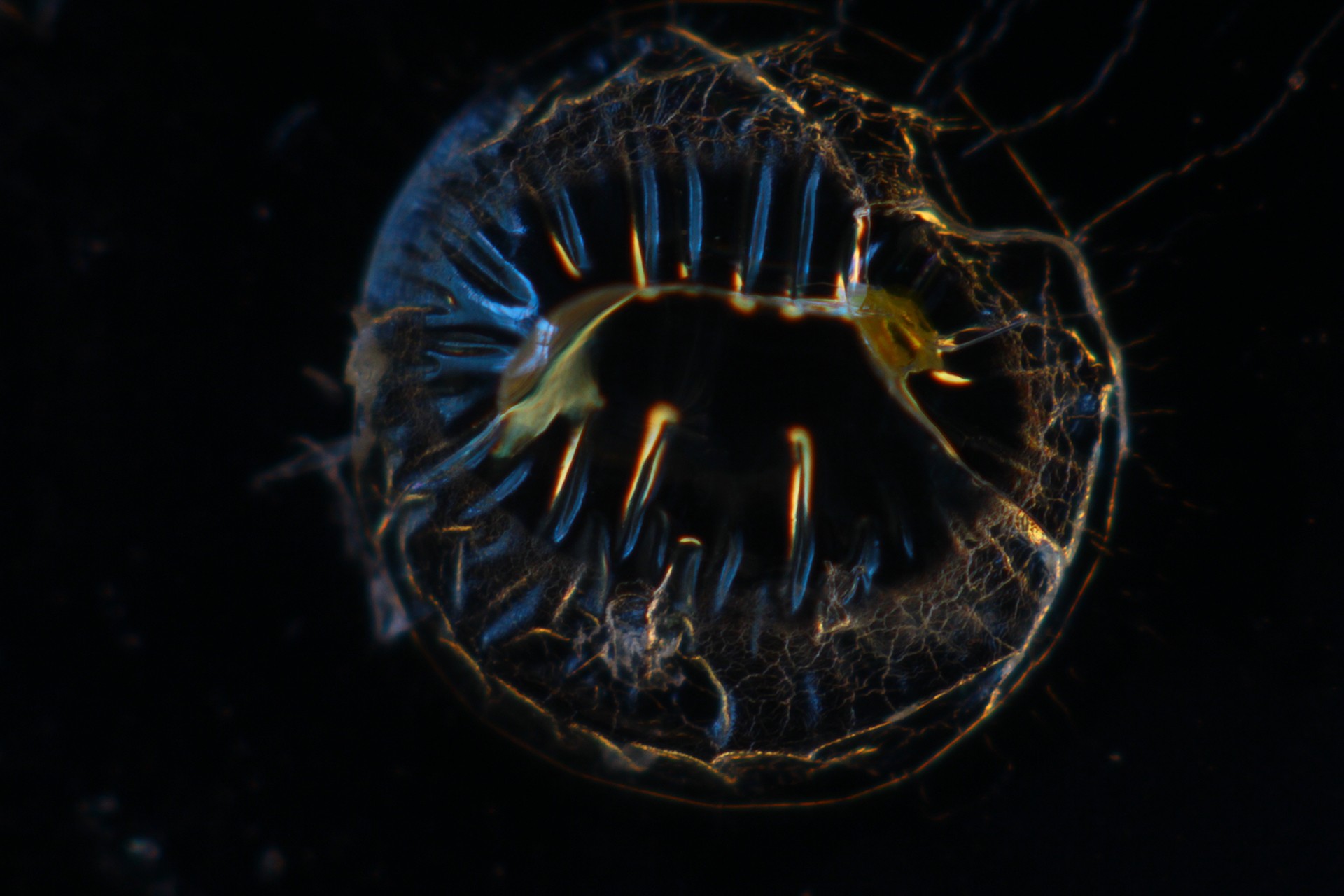
Remnant of an egg capsule just after a juvenile pond snail (Lymnaea stagnalis) has emerged from it. When observed under a dark field of a compound microscope, the defracting and refracting light gave these brilliant blue and golden hues to the otherwise transparent, empty egg capsule. (As seen under a compound microscope and magnified 10x.)
Truly, I can't think of anything more exciting than the process by which a single cell develops into an embryo and finally, into a complete organism.